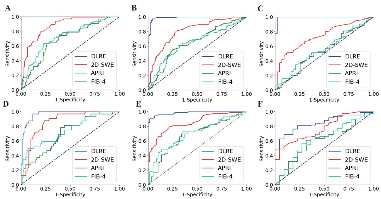
Release date: 2018-05-03 Image source: 16sucai This article was reprinted from the "Automation Institute", the original title: a new breakthrough in the clinical diagnosis of hepatitis B fibrosis. China has nearly 100 million carriers of hepatitis B virus and accounts for more than 50% of chronic hepatitis B patients worldwide. This has long been a serious public health problem and even a social problem in China. Liver fibrosis is a pathological manifestation of chronic hepatitis B patients with cirrhosis and liver cancer. Accurate staging diagnosis is an important basis for clinical surveillance, treatment decision-making and prognosis evaluation. However, clinical staging of liver fibrosis depends on liver biopsy. The method not only has physical trauma to the patient, but also has obvious side effects, and is difficult to re-use, and cannot monitor the development of the patient's condition in a long-term and dynamic manner. Therefore, the clinical diagnosis and treatment of hepatitis B patients has been seeking non-invasive imaging methods to achieve accurate staging of liver fibrosis. In response to this clinical challenge, the Key Laboratory of Molecular Imaging of the Chinese Academy of Sciences has applied a number of technological innovations in the field of imaging omics to the medical imaging big data artificial intelligence analysis technology based on deep learning, which is applied to computer-aided diagnosis of ultrasound elastography. Through the customized design of related models and algorithms, it is possible to diagnose the degree of liver fibrosis in patients based on ultrasound elastic images, non-invasive and intelligent staging, thus assisting doctors to realize personalized treatment decisions for patients with hepatitis B. The related method was led by the Third Affiliated Hospital of Sun Yat-sen University and the General Hospital of the People's Liberation Army, and carried out multi-center, prospective clinical trial verification. A total of 398 patients with hepatitis B were enrolled in 12 hospitals across the country, and about 2000 ultrasound images were collected. The corresponding liver biopsy results were used as the gold standard for imaging ensemble artificial intelligence diagnosis, conventional elastography diagnosis and clinical Serological diagnosis of three methods for the comparison of large data accuracy of hepatitis B fibrosis staging. The statistical results show that the imaging ensemble method has a remarkable accuracy of 97%-100% in the non-invasive diagnosis of liver fibrosis, achieving the same diagnostic efficiency as the invasive liver biopsy. Compared with conventional ultrasound elastography, the accuracy is increased by more than 15% on average; compared with serological diagnosis, the accuracy is increased by more than 30% on average (see the figure below). In addition, the study shows that with the gradual increase of sample size, the diagnostic accuracy of imaging ensemble artificial intelligence will gradually improve, but the accuracy of ultrasound elastography and serological diagnosis can not be improved after peaking. International EFSUMB Clinical Guidelines Recommendation: For individual patients, it is recommended to extract 3 elastic patterns for diagnosis. This test proves that, according to the guidelines, the imaging ensemble method has significantly exceeded the diagnostic accuracy of conventional elastography; if a single patient extracts 5 elastic images, the difference in imaging diagnostics and elastography diagnostic accuracy will further widen. This result demonstrates the significant advantages and strong potential of artificial intelligence to aid clinical diagnosis. Finally, the study also shows that the imaging omics method is extremely robust. Although the hospitals participating in the trial were located in different regions of China, there were no statistical differences in the diagnostic accuracy of multiple imaging ensemble models constructed by artificial intelligence technology to randomly select patients from different hospitals. This provides a solid theoretical foundation and clinical data for the nationwide promotion of the imaging omics method and the true integration into daily clinical diagnostic protocols. Wang Kun, deputy researcher of the Key Laboratory of Molecular Imaging of the Chinese Academy of Sciences, was the first author. Zheng Rongqin, director of the Department of Ultrasound, the Third Affiliated Hospital of Sun Yat-sen University, Tian Jie, researcher of the Key Laboratory of Molecular Imaging of the Chinese Academy of Sciences, and Liang Ping, director of the interventional ultrasound department of the General Hospital of the Chinese People's Liberation Army. For the co-directed author, published in the clinical journal Gut (2018, DOI: 10.1136/gutjnl -2018-316204). Imaging group artificial intelligence (DLRE), ultrasound elastography (2D-SWE) and serological testing (APRI, FIB4) three methods for cirrhosis (Figures A and D), severe liver fibrosis (Figures B and E), Comparison of ROC curves for staging diagnostic efficacy of significant liver fibrosis (Figures C and F) Source: Automation Research Institute Frozen Octopus Frozen octopus ZHEJIANG EVERNEW SEAFOOD CO.,LTD , https://www.evernewseafood.com
Chinese Academy of Sciences: New breakthrough in clinical intelligent diagnosis of hepatitis B fibrosis
Prev Article
High-yield fertilization peanuts