Nature: Revealing the melatonin receptor structure, making sleep better is no longer a dream April 30, 2019 Source: Academic Jingwei The name of melatonin is not unfamiliar to many people. This natural hormone regulates the body's biological clock, so many people turn to over-the-counter melatonin supplements when trying to improve insomnia or jet lag. Scientists hope to develop safer, more effective, and specific drugs for the melatonin pathway to help treat sleep disorders. However, the mechanism by which melatonin acts on receptors has always been like fog, making it difficult to design drugs. The moment to see the clouds is finally here! Recently, in the two papers of the top academic journal Nature, the international team led by famous structural biologist and Professor Vadim Cherezov of the University of Southern California used X-ray free electron laser (XFEL) to reveal the first two humans. The three-dimensional structure of the melatonin receptor. "We want to provide structural information to other researchers to design new drug molecules or to study mutations in these receptors in patients," said co-reporter Professor Cherezov. Melatonin, an important hormone that regulates the circadian clock In each of our brains, there is a structure called the "pineal body" that makes and secretes melatonin. Ideally, as the sun rises and falls, the sky is bright and dark, and the eyes and brain perceive changes in natural light, which will cause the level of melatonin produced by the pineal gland to change, so we will be sleepy after the night falls. When the sky is bright, Xin is waking up. However, modern society's long-distance travel across time zones, day and night work arrangements, and almost 24 hours of uninterrupted exposure to the social network under the artificial blue light source, our body timekeepers are inevitably into chaos. The consequences of disturbing the circadian rhythm should not be underestimated. It can lead to various diseases such as mental, metabolic, and tumor. Many drug developments for insomnia, abnormal circadian rhythms, and mood disorders target two receptors for melatonin. There are two types of melatonin-binding receptors, MT1 and MT2. Past studies have found that these two melatonin receptors are found in many parts of the body, including the brain, retina, cardiovascular system, liver, kidney, spleen, and intestine. The widespread distribution means that the application or lack of melatonin can affect multiple body functions. Although past studies have taught scientists that MT1 receptors play an important role in controlling rhythm, MT2 receptors are closely related to the cyclical activity of melatonin in vivo. However, without knowing the more specific differences between the two receptor proteins, it is difficult to design drugs that selectively target the MT1 receptor without affecting MT2. G protein coupled receptor (GPCR), the "darling" of drug development Melatonin receptors belong to a class of transmembrane proteins called G-protein coupled receptors (GPCRs). There are about 800 GPCRs known in human cells. They are responsible for transmitting signals on the cell surface and play an important role in the physiological and pathological processes of cells. They are considered to be one of the most important drug therapeutic targets. But depicting the three-dimensional structure of GPCR protein molecules has always been a challenge for biophysicists. X-ray crystallography is a method often used by scientists. This method often requires the protein to grow a large enough crystal to obtain a high-resolution image structure. In this two papers, scientists have used a unique approach to solve the problem of growing crystals and collecting X-ray diffraction data. The expressed purified receptor is placed in a membranous gel that grows in the membrane environment by the gel-supporting crystal. The researchers then used a special syringe to allow the microcrystals to form a stream of fine crystals that were scanned with X-rays. Although the size of the obtained crystal is small, the X-ray of the Stanford Linear Accelerator Coherent Light Source (LCLS) is beneficial to its characteristics of ultra-high brightness and femtosecond pulse. The researchers avoided the problem of radiation damage of the crystal under the light source. Hundreds of thousands of scattering images into the crystal to determine the three-dimensional structure of the receptor. Using the same method, the researchers also tested the structure of dozens of mutant receptors to deepen their understanding of the receptor's working mechanism. The high-resolution three-dimensional structure shows that both MT1 and MT2 contain narrow channels that only allow melatonin to bind. A more interesting finding is that by comparing these two structurally similar receptors, the researchers found that some of the larger compounds appeared to target only the MT1 receptor but not the MT2 receptor, which allowed the design of drugs that selectively target MT1. There is a basis on which to base. “Comparing the three-dimensional structure of MT1 and MT2, we can more clearly distinguish the structural differences unique to the two receptors and distinguish their different roles in the circadian clock. Knowing this, we should design a drug that binds only one receptor. Molecules will be easier. This specific combination is important to minimize adverse reactions," said Wei Liu, one of the co-authors of the MT1 structure, and Arizona State University. Worldwide, the number of people affected by sleep disorders is growing, and it is estimated that one out of every three people has experienced short-term insomnia. We expect these two research results to help new drug developers find safer new sleep aids faster and help more people get a sweet sleep. PTFE Coated Guide Wire,Guide Wire,hydrophilic guide wire,medical guide wire Anesthesia Medical Co., Ltd. , https://www.jssinoanesthesias.com
â–²Professor Vadim Cherezov, co-author of the two studies (Source: University of Southern California official website) 
At this point, structural biologists have brought new insights into the three-dimensional structure of the two melatonin receptors. 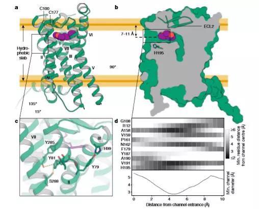
â–² Structural features of melatonin receptor MT1 (Source: Nature) 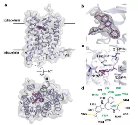
â–² Structural characteristics of melatonin receptor MT2 (Source: Nature) 
â–²The increased expression of nocturnal melatonin (the constellation in the night) and the high affinity of the receptor molecule promotes our sleep (Source: Bridge Institute at USC Michelson Center for Convergent Bioscience, author Yekaterina Kadyshevskaya)
Nature: Revealing the melatonin receptor structure, making sleep better is no longer a dream
Prev Article
Agricultural Machinery Triangle