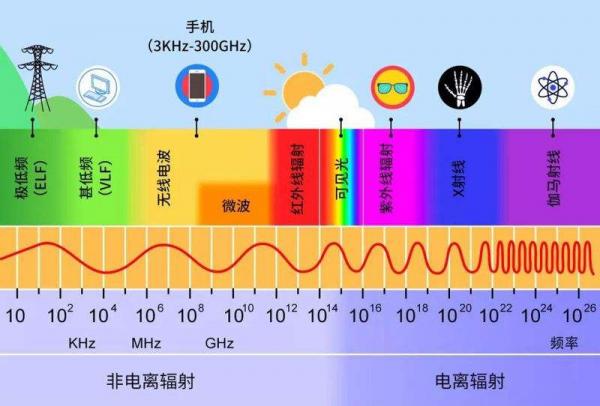
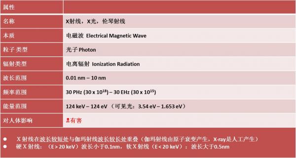
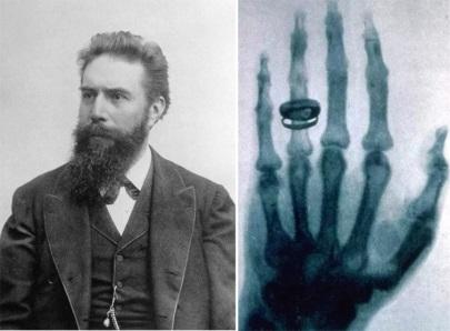
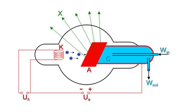
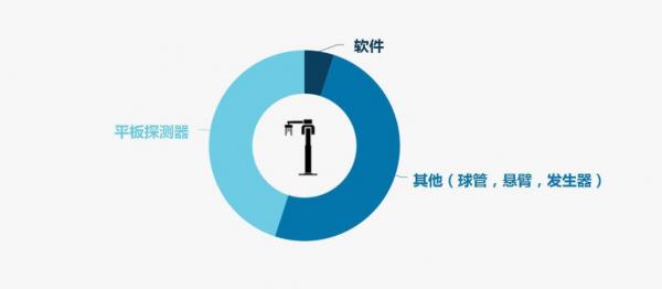
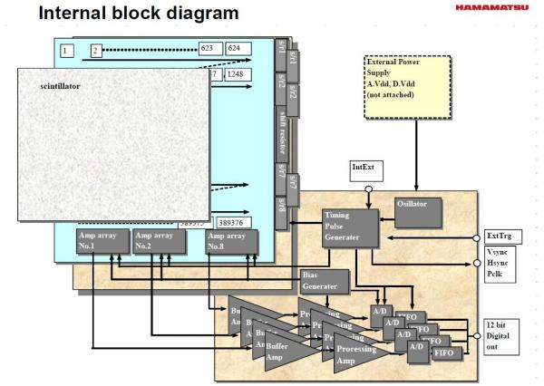
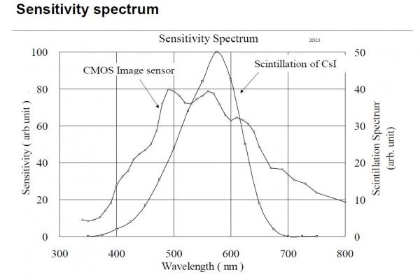
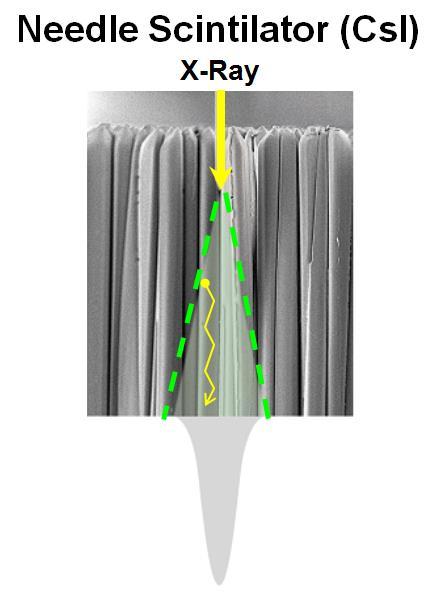
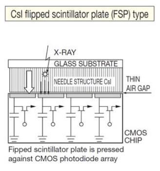
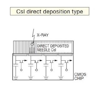
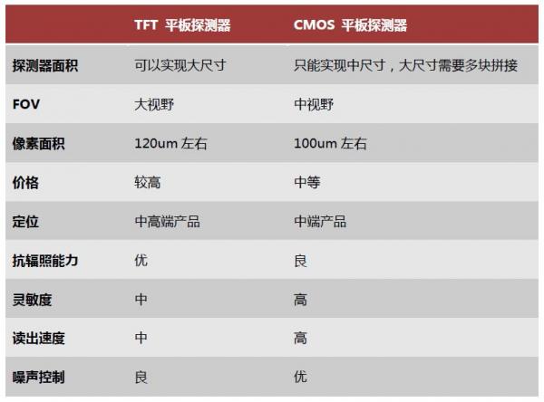
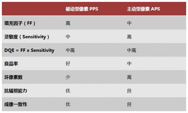
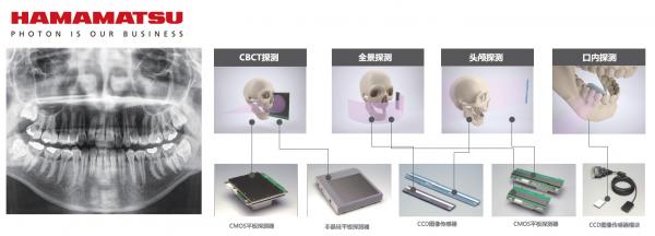
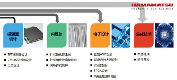
When choosing dental CBCT, if you can understand the equipment itself well, you can undoubtedly help hospitals and dental clinics make purchasing decisions that better suit their needs. But this often requires friends from the medical community to cross the border to understand the issue of optoelectronic technology. why? Because its basic principle is actually X-ray imaging. Understanding it is the most fundamental step in understanding dental CBCT imaging equipment. Today, Xiaobian is here to present some basic knowledge of dental X-ray imaging and how to recognize one of the most important components of dental CBCT - flat panel detector. Everything starts with X-rays Light (radiation) is almost ubiquitous in our lives, in nature, and in the universe. Only a very small part is visible, most of which are unobservable to you and the naked eye. X-ray is one of them. X-rays are high-energy rays. The photon energy is between 124 eV and several hundred keV. It is ionizing radiation and harmful to human body. If the body absorbs excessive X-rays, it will cause substantial damage. There are a lot of X-rays in the universe, but the earth's atmosphere basically separates it. How was it discovered again? The X-ray is also called the Roentgen ray. It was discovered and named in the experiment by the German physicist William Conrad Roentgen in 1895. Roentgen became the first person to win the Nobel Prize in Physics. X-rays are also known as one of the three major discoveries of physics in the late 19th and early 20th centuries, marking the birth of modern physics. In one experiment, Roentgen asked his wife to put her hand on a black paper-wrapped photographic film and irradiate it with X-ray for 15 minutes. After development, the first human X-ray was obtained. This historic photograph also shows that humans can use X-rays to see through the skin. Roentin and Roentgen's palm X-ray The object that refers to the black dragon winter is the wedding ring of Mrs. Roentgen. X-ray penetration is extremely strong. Because different tissues of the human body absorb X-rays differently, evenly distributed X-rays quickly penetrate human tissue, and its uneven distribution is actually the projection of human tissue. Through continuous technological development, X-rays are now widely used in medical imaging, and of course the dental images we are talking about today. How did X-rays in dental images come about? Currently in dental applications, X-ray vacuum tubes are used to generate X-rays. The X-ray tube is a vacuum diode that operates at high voltages. It contains two electrodes: one is a filament for emitting electrons as a cathode (K); the other is a target for receiving electron bombardment as an anode (A). Both poles are sealed in a high vacuum glass or ceramic enclosure. In the parameters of dental X-ray imaging equipment, "tube current" and "tube voltage" are often visible. Here is a small series to explain: The X-ray tube power supply portion includes at least one low voltage power source (Uh) for heating the filament, and a high voltage generator (Ua) for applying a high voltage to the two electrodes. When the tungsten wire passes through enough current to generate an electron cloud, and a sufficient voltage (kV level) is applied between the anode and the cathode (tube voltage), the electron cloud is pulled toward the anode. At this time, the electron hits the tungsten target (A) in a high-energy high-speed state, and the high-speed electron reaches the target surface, and the motion is suddenly blocked, and a small part of the kinetic energy is converted into radiant energy, which is emitted in the form of X-ray. Changing the filament current (tube current) can change the temperature of the filament and the amount of electron emission, thereby changing the tube current and X-ray intensity. Changing the X-ray excitation potential or using a different target can change the energy of the incident X-ray. The higher the tube voltage (keV), the stronger the ability of X-rays to penetrate an object. The tube voltage setting should not be too high or too low. When the tube voltage is too high, most of the X-rays will directly pass through the object. The detector receives the signal and has nothing to do with the object. When the tube voltage is too low, most of the X-rays are absorbed by the object, and the detector receives no. Go to the information about the subject. The larger the tube current (mA), the greater the flow of X-rays and the stronger the signal received by the detector. From the imaging point of view, the larger the tube current, the better, but the larger the tube current, the greater the radiation dose received by the patient, so the smaller the tube current, the better, on the basis of imaging. Due to the high-energy electron bombardment, the X-ray tube operates at a high temperature and requires forced cooling of the anode target. Although X-ray tubes produce X-rays with very low energy efficiency, they are still the most practical X-ray generating devices and have been widely used in X-ray instruments. At present, medical applications mainly include diagnostic X-ray tubes and therapeutic X-ray tubes. The key to dental CBCT imaging: flat panel detector In imaging, in addition to the light source, another essential component is the photodetector. And used in dental CBCT, I think everyone knows about X-ray flat panel detectors. For dental CBCT, flat panel detectors are a central factor affecting image quality. At the same time, from the cost point of view, X-ray flat panel detectors usually account for 1/2-1/3 of the cost of dental CBCT, which fully reflects its importance in the equipment. Therefore, when purchasing a dental CBCT machine, the brand and technical parameters of the flat panel detector are the main points of attention. The proportion of each hardware and software in dental CBCT For X-ray imaging, indirect imaging is currently mostly used, as shown in the following figure (pictured Hamamatsu Dental Flat Panel Detector). We can see that the interior of the flat panel detector mainly contains: Scintillator (gray) Detector (blue) Readout circuit (yellow) Flat panel detector (Hamamatsu Dental Flat Panel Detector) Simply put, the scintillator is responsible for converting X-rays into visible light (X-rays are difficult to detect directly by the detector). The detector is responsible for reading visible light information and converting it into electrical signals. Finally, the readout circuit transmits the electrical signals to the PC. . Here, we focus on the two most critical parts that directly determine the performance of flat panel detectors: scintillators and detectors. 1, scintillator Simply put, a scintillator is a light converter that converts X-rays into visible light that is easily detected. There are many types of scintillators, and currently CsI (geranium iodide scintillator) is mainly used in dental X-rays. The luminescence spectrum of the cesium iodide scintillator is almost perfectly coincident with the absorption spectrum of the CMOS image sensor, and the peaks of both curves fall within the spectral range of 500-600 nm. At present, the highest resolution in the cesium iodide scintillator is acicular cesium iodide. The needle-like structure is similar to the light guide. When the visible light is output, the light is more concentrated, the brightness is stronger, and the resolution is higher. Needle-shaped cesium iodide scintillator The scintillator needs to be coupled to the detector to be effective, and the coupling is mainly divided into two types: 1. Substrate pressing (coupling) type 2. Direct deposition type The substrate is pressed (coupled) type scintillator, which is a substrate on which a scintillator is grown, and then the substrate is placed thereon, and the scintillator is buckled downward on the flat panel detector. There is a thin gap between the needle-shaped cesium iodide and the flat panel detector in the pressing process, and there is residual air in the inside; in the coupling process, there is a glue layer in the gap, and the substrate pressing (coupling) type scintillator is inevitably It is the interception absorption of X-ray by the substrate, and the refraction and scattering of the converted light by the air layer or the glue layer. This affects the intensity and resolution of the scintillator. However, this type of scintillator is relatively difficult to produce and relatively low in cost. Directly deposited acicular yttrium iodide scintillator, no adhesive, no additional substrate, direct evaporation on flat panel detectors (CMOS and TFT plates) by professional equipment, flashing needle-shaped cesium iodide The body grows on the surface of the flat panel detector. This coupling method, because there is no additional substrate and adhesive or air layer, the incident X-ray and emitted visible light loss is small, the luminous intensity is high, the illumination is concentrated, the resolution is high, and it can be irradiated under low dose X-ray. Produces good image quality. The direct deposition type acicular cesium iodide scintillator is the highest standard for the scintillation process used in X-ray flat panel detectors. This coupling method is technically difficult and costly. At present, Hamamatsu's flat-panel detectors are all directly deposited. The Hamamatsu company has mastered the production process of direct-deposited needle-shaped cesium iodide, which can produce high-quality direct-deposited needle-shaped cesium iodide. Scintillator. 2, detector The detector is located below the scintillator and is used to receive the visible light emitted by the scintillator and convert the optical signal into an electrical signal, which is a key step in photoelectric conversion. There are currently two detectors on the market for dental CBCT imaging: CMOS flat panel detector TFT flat panel detector (also known as amorphous silicon flat panel detector) At present, the pros and cons of these two types of detectors can be said to be different. As a Hamamatsu with two detectors at the same time, let's share the actual comparison between the two: As can be seen from the table, the advantages of TFT flat panel detectors are size and field of view, while the advantages of CMOS flat panel detectors are sensitivity, resolution and readout speed. CMOS panels have an advantage in performance without the need for splicing. However, in applications requiring "large size", the spliced ​​CMOS panel will have gaps and failed pixel lines at the splicing, which will result in partial image loss, which needs to be corrected by software later. The TFT tablet does not have this problem. At present, Hamamatsu's mid-view panels are all CMOS panels. The medium-large field of view and the large-view panel are all TFT panels, which can fully exploit the advantages of each process and meet the application needs of different scenarios. Next, let's take a look at another key point in the detector that we must understand - the pixel structure. At present, the pixel structure of flat panel detectors is generally divided into two types: Passive PPS: The entire area of ​​the pixel is used to detect the optical signal, and the pixel does not contain an amplifier; Active Pixel APS: Each pixel contains a photodetector and amplifier. For the flat panel detector products currently on the market, the TFT flat panel detector uses passive pixel PPS, while the CMOS flat panel detector has two pixel structures. At present, the X-ray dose required for dental CBCT imaging is between 20uSv and 50uSv. At this dose, the imaging quality of APS type CMOS flat panel detector, PPS type CMOS flat panel detector and TFT flat panel detector is almost the same. Through the above sharing, we should have a better understanding of the flat panel detector for dental CBCT. Here, Xiaobian will make a small summary of knowledge for everyone: Knock on the blackboard! Focus on ~ · TFT flat panel detectors and CMOS flat panel detectors have their own strengths. The TFT process is mostly used to manufacture large-area flat panel detectors, which are mostly used in high-end seated dental CBCT machines. The CMOS process is mostly used to manufacture mid-field flat panel detectors, which are mostly used in dental 3-in-1 models (but there are also some high-end 3). -in-1 model, using a large field of view flat panel detector in TFT); · Directly deposited acicular cesium iodide TFT and CMOS flat panel detectors are the highest quality products in the dental flat panel detector industry; ·Spliced ​​CMOS flat panel detectors will have obvious bad lines, relying heavily on algorithmic compensation; · Each pixel in the APS type CMOS detector contains an amplifier. X-rays that are not completely absorbed by the scintillator easily cause damage to the amplifier, causing pixel failure, and its radiation resistance is not as good as that of the PPS type CMOS flat panel detector. Ok, I hope that the above content will help you to know more about dental X-ray imaging, especially dental CBCT equipment. As a photovoltaic company with more than 60 years, Hamamatsu has a history of 24 years in the development and production of flat panel detectors. Hamamatsu flat panel detectors continue to provide excellent products for the world's leading dental imaging companies by mastering the core technologies - detector design, scintillator production, electronic design and product integration. It sells tens of thousands of dental flat panel detectors every year, and its global market share exceeds 50%. We also hope to provide better possibilities for the development of dental imaging by continuously improving our own technology. The world-renowned dental imaging equipment manufacturers (parts) served by Hamamatsu: Sirona, KaVo, Ray, Morita, etc. Hamamatsu offers a full line of detector solutions for dental imaging applications Hamamatsu flat panel detector core technology, in addition to dental applications, Hamamatsu flat panel detectors are also widely used in industrial non-destructive testing.
[Sample requirements] Vtm Sampling Tube With Swab,Disposable Collection Tube Of Virus Samples,Hi Media Vtm Kit,Amylase Sample Collection Tube Jilin Sinoscience Technology Co. LTD , https://www.contoryinstruments.com

The collected nasopharyngeal swab samples should be transported at 2°C to 8°C and sent for inspection immediately, and the sample delivery and storage time should not exceed 48 hours.
[Testing method]
1. Before sampling, mark the relevant sample information on the label of the sampling tube.
2. According to different sampling requirements, use a sampling swab to sample in the nasopharynx.
3. The specific sampling methods are as follows:
a) Nasal swab: Gently insert the swab head into the nasal palate, stay for a while and then slowly turn to exit. Wipe the other nostril with another swab, immerse the swab head in the sampling solution, and discard the tail.
b) Pharyngeal swab: Wipe bilateral pharyngeal tonsils and posterior pharyngeal wall with a swab, also immerse the swab head in the sampling solution, and discard the tail.
4. Quickly put the swab into the sampling tube.
5. Break the part of the sampling swab higher than the sampling tube, and tighten the tube cover.
6. Freshly collected clinical specimens should be transported to the laboratory within 48 hours at 2°C to 8°C.
[Explanation of test results]
After the sample is collected, the sampling solution turns slightly yellow, which will not affect the nucleic acid test result.
[Limitations of the test method]
1. For samples that are seriously contaminated due to improper storage after collection, the final test results will be affected.
2. If the sample is not stored at the specified temperature, the final test result will be affected.
It turns out that the key to affecting dental CBCT imaging is here!